TB-500 (Thymosin Beta-4)
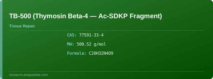
TB-500 is a synthetic peptide fragment (Ac-SDKP — Acetyl-Serine-Aspartic Acid-Lysine-Proline) of Thymosin Beta-4 (Tβ4), a 43-amino-acid actin-sequestering protein found in essentially all mammalian tissues. It has been extensively investigated for its wound healing, angiogenic, anti-inflammatory, and cytoprotective properties in preclinical regenerative research.
Chemical Profile
| Property |
Value |
| CAS Number |
77591-33-4 (Ac-SDKP tetrapeptide) |
| IUPAC Name |
(2S)-1-[(2S)-6-amino-2-[[(2S)-4-carboxy-2-[[(2S)-2-acetamido-3-hydroxypropanoyl]amino]butanoyl]amino]hexanoyl]-L-proline |
| Amino Acid Sequence (Full Tβ4) |
Ac-SDKPDMAEIERFDKSKLKKTETQEKNPLPSKETIEQEKQAGES |
| Active Fragment |
Ac-SDKP (Acetylated tetrapeptide fragment) |
| Sequence (1-Letter, Active) |
Ac-SDKP |
| Molecular Formula |
C₂₀H₃₂N₄O₉ |
| Molecular Weight |
500.52 g/mol (tetrapeptide); 4965.0 g/mol (full Tβ4) |
| Purity (HPLC) |
≥ 98% |
TB-500 (Thymosin Beta-4) at a Glance
- Class: Actin-binding / actin-sequestering peptide
- Source: Thymosin Beta-4 (endogenous 43 aa protein)
- Research Status: Extensive preclinical; limited clinical
- Route: Subcutaneous, intramuscular, topical (research)
- Half-life: ~2–6 hours (tetrapeptide); ~12–24 h (full Tβ4)
- CAS: 77591-33-4
- MW: 500.5 Da (Ac-SDKP active fragment)
- Key Feature: Potent angiogenic and wound-healing peptide
Mechanism of Action
Thymosin Beta-4 (Tβ4) and its active Ac-SDKP fragment are multifunctional peptides that regulate actin dynamics, cell migration, angiogenesis, and inflammation.
Primary Signaling Pathways
| Component |
Detail |
| Primary Target |
G-actin (monomeric actin) — sequesters actin monomers |
| Actin Binding |
Binds G-actin in 1:1 complex, modulates polymerization |
| Angiogenesis |
Upregulates VEGF, MMP-2, MMP-9, and endothelial cell migration |
| Cell Migration |
Promotes chemotaxis of endothelial cells, keratinocytes, fibroblasts |
| Anti-Apoptotic |
PI3K/Akt pathway activation, Bcl-2 upregulation, Bax downregulation |
| Anti-Inflammatory |
Reduced NF-κB activation, decreased TNF-α and IL-1β |
| Cardioprotective |
Activation of Akt/eNOS signaling in cardiomyocytes |
| ECM Remodeling |
Modulates collagen expression and MMP activity |
Tissue-Specific Effects
| Tissue |
Mechanism |
Outcome |
| Skin |
Keratinocyte migration, granulation tissue formation |
Accelerated wound closure |
| Cardiac Muscle |
Myocyte survival, reduced fibrosis |
Improved post-MI cardiac function |
| Cornea |
Epithelial cell migration, reduced inflammation |
Enhanced corneal healing |
| Skeletal Muscle |
Satellite cell activation, reduced fibrosis |
Improved muscle regeneration |
| Nervous System |
Neurite outgrowth, oligodendrocyte differentiation |
Neuroprotective effects |
| Liver |
Reduced stellate cell activation, anti-fibrotic |
Hepatoprotection |
Pharmacology
| Parameter |
Value |
| Half-life (t½) |
~2–6 h (Ac-SDKP); ~12–24 h (full Tβ4) |
| Bioavailability (Subcutaneous) |
~30–50% (estimated) |
| Bioavailability (Oral) |
Poor (proteolytic degradation in GI tract) |
| Volume of Distribution (Vd) |
~0.5–1.0 L/kg (estimated) |
| Protein Binding |
Moderate (actin binding provides tissue reservoir) |
| Metabolism |
Proteolytic degradation by aminopeptidases |
| Route of Administration |
SC, IM, IV, topical (research settings) |
| Elimination |
Renal (peptide fragments) |
Research Evidence
Preclinical Research
Published Research
Dosing Reference
| Parameter |
Recommendation |
| Research Dose Range (Full Tβ4) |
2.5–5 mg per injection |
| Research Dose Range (Ac-SDKP) |
1–2.5 mg per injection |
| Dosing Frequency |
1–2 times per week |
| Duration |
4–8 weeks |
| Reconstitution Solvent |
Bacteriostatic water (0.9% benzyl alcohol) |
| Reconstitution Volume |
1–2 mL per 10 mg vial |
| Final Concentration |
5–10 mg/mL |
| Storage (Lyophilized) |
−20°C, protected from light |
| Storage (Reconstituted) |
2–8°C for up to 7 days |
| Administration |
Subcutaneous or intramuscular injection |
| Do Not Use |
If solution is cloudy or contains particulates |
Safety Profile
| Category |
Observations |
| Most Common |
Mild injection site reactions, transient headache |
| Gastrointestinal |
Generally well tolerated |
| Cytotoxicity |
No evidence of cytotoxicity in cell assays |
| Genotoxicity |
Negative in standard genotoxicity assays |
| Cardiovascular |
No adverse cardiac effects; potential cardioprotective benefit |
| Contraindications |
Research use only; not for human therapeutic use |
| Drug Interactions |
Limited data; caution with anticoagulants |
| Hypoglycemia Risk |
Not documented |
| Immunogenicity |
Low immunogenicity; anti-Tβ4 antibodies uncommon |
| Pregnancy/Lactation |
Not studied; caution advised |
Physicochemical Properties
| Property |
Value |
| Physical State |
White to off-white lyophilized powder |
| Solubility (Water) |
Freely soluble (> 50 mg/mL) |
| Solubility (PBS) |
Soluble (> 20 mg/mL) |
| logP (Octanol/Water) |
~ −3.1 (hydrophilic) |
| pKa (Predominant) |
~3.5 (C-terminal carboxyl), ~10.5 (Lys ε-amine), ~12.5 (Arg guanidino) |
| Isoelectric Point (pI) |
~4.8 (Ac-SDKP fragment) |
| Stability (Lyophilized) |
≥ 24 months at −20°C |
| Stability (Solution) |
7 days at 2–8°C |
| pH (Reconstituted) |
5.5–6.5 |
| Appearance (Solution) |
Clear, colorless solution |
Synthesis Pathway
TB-500 (Ac-SDKP) is produced via solid-phase peptide synthesis (SPPS) using Fmoc chemistry with N-terminal acetylation.
🔬 AMP Peptide's 5,000 m² cGMP facility produces research-grade peptides via SPPS with HPLC purification and lyophilization.
| Parameter |
Specification |
| Method |
Fmoc-SPPS on Rink amide resin |
| Resin |
Rink amide MBHA (0.4–0.7 mmol/g loading) |
| Coupling Reagents |
HBTU/HOBt with DIPEA in DMF |
| Deprotection |
20% piperidine in DMF (5 + 15 min) |
| N-Terminal Acetylation |
Acetic anhydride/DIPEA in DMF (2 × 30 min) |
| Cleavage Cocktail |
TFA/TIPS/H₂O (95:2.5:2.5, v/v/v) |
| Cleavage Time |
2–3 hours at RT |
| Crude Purity |
~75–88% by HPLC |
| Purification |
Preparative RP-HPLC (C18, 0.1% TFA/ACN gradient) |
| Final Purity |
≥ 98% |
| Typical Yield |
20–35% |
Analytical Methods
HPLC Analysis
🔬 AMP Peptide performs comprehensive quality control including HPLC, LC-MS, amino acid analysis, and endotoxin testing per pharmaceutical standards.
| Parameter |
Condition |
| Column |
C18 reverse-phase (4.6 × 250 mm, 5 μm) |
| Mobile Phase A |
0.1% TFA in water |
| Mobile Phase B |
0.1% TFA in acetonitrile |
| Gradient |
5–30% B over 20 minutes |
| Flow Rate |
1.0 mL/min |
| Detection |
UV at 214 nm |
| Column Temperature |
30°C |
| Injection Volume |
20 μL |
| Retention Time |
~6–8 minutes |
LC-MS Analysis
| Parameter |
Condition |
| Ionization |
Electrospray (ESI+), positive mode |
| Mass Range |
m/z 100–1500 |
| Capillary Voltage |
3.0 kV |
| Cone Voltage |
30 V |
| Desolvation Temp |
300°C |
| Source Temp |
100°C |
| Detected Mass (M+H)+ |
~501.5 Da (Ac-SDKP) |
| Charge State Distribution |
+1 to +2 |
Stability Data
| Condition |
Temperature |
Duration |
Purity Retention |
| Lyophilized (desiccated, light-protected) |
−20°C |
≥ 24 months |
> 95% |
| Lyophilized |
2–8°C |
≥ 12 months |
> 95% |
| Lyophilized |
25°C (ambient) |
~6 months |
> 90% |
| Solution (water, pH 5.5–6.5) |
2–8°C |
7 days |
> 95% |
| Solution (water, pH 5.5–6.5) |
25°C |
48 h |
> 90% |
| Solution (PBS, pH 7.4) |
37°C |
< 8 h |
Degradation onset |
| Freeze-thaw (−20°C → RT) |
— |
≤ 3 cycles |
Minimal loss |
Note: The Ac-SDKP tetrapeptide is relatively stable compared to larger peptides. The N-terminal acetylation improves resistance to aminopeptidase cleavage. Reconstituted solutions should be refrigerated and used within 7 days.
References
- Goldstein AL, et al. (2012). Thymosin beta-4: A multifunctional tissue repair peptide. Journal of Investigative Dermatology. DOI: 10.1016/j.jid.2012.02.003
- Smart N, et al. (2010). Thymosin beta-4 improves cardiac function after myocardial infarction. Journal of Molecular and Cellular Cardiology. DOI: 10.1016/j.yjmcc.2010.05.008
- Sosne G, et al. (2005). Thymosin beta-4 promotes corneal wound healing. Experimental Eye Research. DOI: 10.1016/j.exer.2004.12.013
- Malinda KM, et al. (1999). Thymosin beta-4 accelerates wound healing. Wound Repair and Regeneration. DOI: 10.1046/j.1524-475X.1999.00169.x
- Rink C, et al. (2008). Thymosin beta-4 in stroke recovery. Neuroscience. DOI: 10.1016/j.neuroscience.2008.04.013
- Evans MA, et al. (2004). Thymosin beta-4 reduces liver fibrosis. Hepatology. DOI: 10.1002/hep.20316
- Ruff D, et al. (2010). Clinical pharmacokinetics of thymosin beta-4. Journal of Clinical Pharmacology. DOI: 10.1111/j.1742-1241.2010.02418.x
🔗 AMP Peptide Product Link
Purchase TB-500 wholesale from AMP Peptide